


Gross and Microscopic Anatomy of the Stomach(histology).
:: أقسام الكليه :: الفرقه الاولى
صفحة 1 من اصل 1

 Gross and Microscopic Anatomy of the Stomach(histology).
من طرف د/على نور الثلاثاء 22 سبتمبر 2009, 6:22 pm
Gross and Microscopic Anatomy of the Stomach

The
stomach is an expanded section of the digestive tube between the
esophagus and small intestine. It's characteristic shape is shown,
along with terms used to describe the major regions of the stomach. The
right side of the stomach shown above is called the greater curvature
and that on the left the lesser curvature. The most distal and narrow
section of the stomach is termed the pylorus - as food is liquefied in the stomach it passes through the pyloric canal into the small intestine.
The
wall of the stomach is structurally similar to other parts of the
digestive tube, with the exception that the stomach has an extra,
oblique layer of smooth muscle inside the circular layer, which aids in
performance of complex grinding motions.
===================
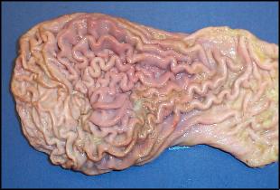
In
the empty state, the stomach is contracted and its mucosa and submucosa
are thrown up into distinct folds called rugae; when distended with
food, the rugae are "ironed out" and flat. The image to the right shows
rugae on the surface of a dog's stomach.

Within
the stomach there is an abrupt transition from stratified squamous
epithelium extending from the esophagus to a columnar epithelium
dedicated to secretion. In most species, this transition is very close
to the esophageal orifice, but in some, particular horses and rodents,
stratified squamous cells line much of the fundus and part of the body.
The
image to the right is of the mucosal surface of an equine stomach
showing esophageal epithelium (top) and glandular epithelium (bottom).
The creatures attached to the surface are bots, larval forms of Gasterophilus
=============================================
Four major types of secretory epithelial cells cover the surface of the stomach and extend down into gastric pits and glands:
are differences in the distribution of these cell types among regions
of the stomach - for example, parietal cells are abundant in the glands
of the body, but virtually absent in pyloric glands. The micrograph to
the right shows a gastric pit invaginating into the mucosa (fundic
region of a raccoon stomach). Notice that all the surface cells and the
cells in the neck of the pit are foamy in appearance - these are the
mucous cells. The other cell types are farther down in the pit and, in
this image, difficult to distinguish.

[ندعوك للتسجيل في المنتدى أو التعريف بنفسك لمعاينة هذا الرابط]
Digestive Anatomy in Ruminants
The stomach of ruminants has four compartments: the rumen, reticulum, omasum and abomasum, as shown in the following diagram:
The interior surface of
the rumen forms numerous papillae
that vary in shape and size from short and pointed to long and foliate.
=================================================

Reticular
epithelium is thrown into folds that form polygonal cells that give it
a reticular, honey-combed appearance</B>. Numerous small papillae
stud the interior floors of these cells.
=======================================

The inside of the omasum
is thrown into broad longitudinal folds or leaves</B> reminiscent
of the pages in a book (a lay term for the omasum is the 'book'). The
omasal folds, which in life are packed with finely ground ingesta, have
been estimated to represent roughly one-third of the total surface area
of the forestomachs.

[ندعوك للتسجيل في المنتدى أو التعريف بنفسك لمعاينة هذا الرابط]
Dynamics of Cranial Digestion
Feed,
water and saliva are delivered to the reticulorumen through the
esophageal orifice. Heavy objects (grain, rocks, nails) fall into the
reticulum, while lighter material (grass, hay) enters the rumen proper.
Added to this mixture are voluminous quantities of gas produced during
fermentation.
Ruminants
produce prodigious quantities of saliva. Published estimates for adult
cows are in the range of 100 to 150 liters of saliva per day! Aside
from its normal lubricating qualities, saliva serves at least two very
important functions in the ruminant:
these materials within the rumen partition into three primary zones
based on their specific gravity. Gas rises to fill the upper regions,
grain and fluid-saturated roughage ("yesterday's hay") sink to the
bottom, and newly arrived roughage floats in a middle layer.

Reticuloruminal Motility
in
orderly pattern of ruminal motility is initiated early in life and,
except for temporary periods of disruption, persists for the lifetime
of the animal. These movements serve to mix the ingesta, aid in
eructation of gas, and propel fluid and fermented foodstuffs into the
omasum. If motility is suppressed for a significant length of time,
ruminal impaction may result. A
cycle of contractions occurs 1 to 3 times per minute. The highest
frequency is seen during feeding, and the lowest when the animal is
resting. Two types of contractions are identified:
Primary contractions
originate in the reticulum and pass caudally around the rumen. This
process involves a wave of contraction followed by a wave of
relaxation, so as parts of the rumen are contracting, other sacs are
dilating.
animation below is based on data collected by radiographing sheep
(Wyburn, 1980) and should impart at least some appreciation of the
complexity of ruminal motility. Although shown much faster than in
life, the major reticuloruminal contractions are timed appropriately.
Note the movements which bring the gas bubble (stippled area) forward
to the esophagus for eructation.

The
stomach is an expanded section of the digestive tube between the
esophagus and small intestine. It's characteristic shape is shown,
along with terms used to describe the major regions of the stomach. The
right side of the stomach shown above is called the greater curvature
and that on the left the lesser curvature. The most distal and narrow
section of the stomach is termed the pylorus - as food is liquefied in the stomach it passes through the pyloric canal into the small intestine.
The
wall of the stomach is structurally similar to other parts of the
digestive tube, with the exception that the stomach has an extra,
oblique layer of smooth muscle inside the circular layer, which aids in
performance of complex grinding motions.
===================
In
the empty state, the stomach is contracted and its mucosa and submucosa
are thrown up into distinct folds called rugae; when distended with
food, the rugae are "ironed out" and flat. The image to the right shows
rugae on the surface of a dog's stomach.
Within
the stomach there is an abrupt transition from stratified squamous
epithelium extending from the esophagus to a columnar epithelium
dedicated to secretion. In most species, this transition is very close
to the esophageal orifice, but in some, particular horses and rodents,
stratified squamous cells line much of the fundus and part of the body.
The
image to the right is of the mucosal surface of an equine stomach
showing esophageal epithelium (top) and glandular epithelium (bottom).
The creatures attached to the surface are bots, larval forms of Gasterophilus
=============================================
Four major types of secretory epithelial cells cover the surface of the stomach and extend down into gastric pits and glands:
- [b]Mucous cells: secrete an alkaline mucus that protects the epithelium against shear stress and acid
- Parietal cells: secrete hydrochloric acid
- Chief cells: secrete pepsin, a proteolytic enzyme
- G cells: secrete the hormone gastrin
are differences in the distribution of these cell types among regions
of the stomach - for example, parietal cells are abundant in the glands
of the body, but virtually absent in pyloric glands. The micrograph to
the right shows a gastric pit invaginating into the mucosa (fundic
region of a raccoon stomach). Notice that all the surface cells and the
cells in the neck of the pit are foamy in appearance - these are the
mucous cells. The other cell types are farther down in the pit and, in
this image, difficult to distinguish.
[ندعوك للتسجيل في المنتدى أو التعريف بنفسك لمعاينة هذا الرابط]
Digestive Anatomy in Ruminants
The stomach of ruminants has four compartments: the rumen, reticulum, omasum and abomasum, as shown in the following diagram:
The interior surface of
the rumen forms numerous papillae
that vary in shape and size from short and pointed to long and foliate.
=================================================
Reticular
epithelium is thrown into folds that form polygonal cells that give it
a reticular, honey-combed appearance</B>. Numerous small papillae
stud the interior floors of these cells.
=======================================
The inside of the omasum
is thrown into broad longitudinal folds or leaves</B> reminiscent
of the pages in a book (a lay term for the omasum is the 'book'). The
omasal folds, which in life are packed with finely ground ingesta, have
been estimated to represent roughly one-third of the total surface area
of the forestomachs.
[ندعوك للتسجيل في المنتدى أو التعريف بنفسك لمعاينة هذا الرابط]
Dynamics of Cranial Digestion
Feed,
water and saliva are delivered to the reticulorumen through the
esophageal orifice. Heavy objects (grain, rocks, nails) fall into the
reticulum, while lighter material (grass, hay) enters the rumen proper.
Added to this mixture are voluminous quantities of gas produced during
fermentation.
Ruminants
produce prodigious quantities of saliva. Published estimates for adult
cows are in the range of 100 to 150 liters of saliva per day! Aside
from its normal lubricating qualities, saliva serves at least two very
important functions in the ruminant:
- provision of fluid for the fermentation vat
- alkaline
buffering - saliva is rich in bicarbonate, which buffers the large
quanitity of acid produced in the rumen and is probably critical for
maintainance of rumen pH.
these materials within the rumen partition into three primary zones
based on their specific gravity. Gas rises to fill the upper regions,
grain and fluid-saturated roughage ("yesterday's hay") sink to the
bottom, and newly arrived roughage floats in a middle layer.
Reticuloruminal Motility
in
orderly pattern of ruminal motility is initiated early in life and,
except for temporary periods of disruption, persists for the lifetime
of the animal. These movements serve to mix the ingesta, aid in
eructation of gas, and propel fluid and fermented foodstuffs into the
omasum. If motility is suppressed for a significant length of time,
ruminal impaction may result. A
cycle of contractions occurs 1 to 3 times per minute. The highest
frequency is seen during feeding, and the lowest when the animal is
resting. Two types of contractions are identified:
Primary contractions
originate in the reticulum and pass caudally around the rumen. This
process involves a wave of contraction followed by a wave of
relaxation, so as parts of the rumen are contracting, other sacs are
dilating.
- Secondary contractions occur in only parts of the rumen and are usually associated with eructation.
animation below is based on data collected by radiographing sheep
(Wyburn, 1980) and should impart at least some appreciation of the
complexity of ruminal motility. Although shown much faster than in
life, the major reticuloruminal contractions are timed appropriately.
Note the movements which bring the gas bubble (stippled area) forward
to the esophagus for eructation.

د/على نور- المـديـر العـــام

- عدد المساهمات : 791
تاريخ التسجيل : 04/03/2009
العمر : 33
الموقع : فى بيتنا -

» wonderful webpage for Biochemistry,Histology and physiology
» wonderful webpage for Biochemistry,Histology and physiology
» CD-Anatomy
» A Colour Atlas of Avian Anatomy
» Pictures Of Anatomy Organ Comparative
» wonderful webpage for Biochemistry,Histology and physiology
» CD-Anatomy
» A Colour Atlas of Avian Anatomy
» Pictures Of Anatomy Organ Comparative
:: أقسام الكليه :: الفرقه الاولى
صفحة 1 من اصل 1
صلاحيات هذا المنتدى:
لاتستطيع الرد على المواضيع في هذا المنتدى